18 Mar How The Eyes Different Parts Power Your Vision
March is Save Your Vision Month, as designated by the American Optometric Association (AOA). This month is an ideal time to increase awareness of the importance of scheduling a comprehensive annual eye examination with your optometrist.
Optometrists diagnose eye diseases through annual eye exams and help you see your best with the right prescription and advice on the optimal type of correction for your lifestyle. Did you know that they can also diagnose other types of disease during an eye exam? For example, in 2014, during comprehensive eye exams, optometrists diagnosed diabetes in 401,000 patients who were not aware they had the disease!
The AOA reports that more than 40 million aging Americans are at risk for reduced vision and potentially even blindness because of age-related diseases. Your optometrist is your first line of defense to prevent reduced vision and blindness. Still, it’s up to you to go in for your annual eye exam so he or she can thoroughly check your eyes for signs of glaucoma, diabetic eye disease, cataracts and age-related macular degeneration. The devastating effects of these diseases can be prevented or slowed with early detection.
Understanding Your Eye
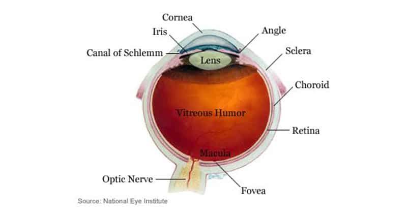
Below, we explore various parts of the eye that your doctor will examine in-depth to make sure your eyes are healthy.
As we identify and describe the different parts of the eye, we’ll also note which problems and diseases occur in them:
Iris. The colored portion of your eye is the iris, composed of fibrovascular tissue. It connects to the tiny muscles that enable your pupils to grow larger or smaller depending on lighting conditions. Diseases of the iris are rare.
Pupil. The pupil is the black tissue at the center of the iris. It is often compared with the aperture of a camera because of its ability to expand and constrict in differing lighting conditions to control the amount of light that enters the eye. This is what enables us to see both in sunny, bright daylight and near-darkness. The reason the pupil appears to be black is that it absorbs most of the light that goes through it.
Sclera. The sclera is “the whites of your eyes.” It is fibrous tissue with collagen in it that serves to protect the eye’s inner structures from damage.
Conjunctiva. This name may be familiar to you because of a disease associated with it: conjunctivitis, an inflammatory condition also known as pinkeye. The conjunctiva is a clear layer covering the sclera (the whites of your eye.) The conjunctiva extends to the inside of your eyelids and produces tears and mucus to lubricate the eyes and keep foreign bodies out.
Cornea. The clear covering over your iris and pupil is called the cornea—the part of the eye that provides most of your optical power. The cornea refracts (bends) light to enable your eyes to focus on objects. Problems related to the cornea can include corneal abrasions, keratoconus, pterygium and astigmatism.
Lens. The lens is a transparent structure within the eye, oblong in shape and curved both outward and inward. It refracts light. Depending on the curvature of your lenses, you may develop astigmatism and have difficulty focusing. Another common problem with the lens as people age is a cataract, the condition in which the lens impairs vision by becoming cloudy and difficult to see through, like a dirty windshield.
Retina. Covering the back surface of the eye, the retina is a sensory membrane. As your eye “sees,” the lens delivers images to the retina, which translates them into electrical signals that the optic nerve delivers to your brain. Diseases and challenges for this area of the eye include retinal detachment, diabetic retinopathy, retinoblastoma and retinitis pigmentosa.
Macula. The center of the retina is called the macula, where your ability to see objects in great detail resides. Aging can sometimes lead to macular degeneration, a disease in which central vision is impaired and patients can no longer see in detail to enjoy daily activities including reading, needlework and watching television.
Aqueous Humor is a thick fluid that provides nutrients to the structures of the eye. This liquid is in an ongoing cycle of draining and being replaced. When patients develop glaucoma, the aqueous humor fluid builds up and creates an uncomfortable feeling of high interocular pressure. Glaucoma can damage your eyesight permanently and cause blindness if left untreated.
Optic Nerve. This nerve connects your eye to the brain and is the pathway by which the retina’s rods and cones send visual signals that are translated by the brain. If the optic nerve isn’t healthy and functioning well, you would lose your ability to see.
Save Your Vision month is the perfect opportunity to call us and schedule an annual eye exam—both for yourself and those you love.


No Comments